The patient is a 59-year-old woman with a history of severe injuries sustained as a nursing student from a patient who attacked her with an oxygen bottle. Her injuries were to the back, head, and knees. The patient has been diagnosed with schizophrenia, depression, and bipolar disorders. She was diagnosed with Systemic Lupus Erythematosus treated with prednisone for 5 years but has not taken prednisone for 1.5 years. After attaining a body weight of 315 pounds she discontinued the prednisone and lost 55 pounds.
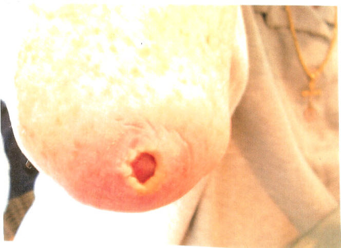
The patient first complained of a pressure ulcer on the right elbow for over a year. The pressure ulcer developed from sleeping in a chair with her elbows on a table. She does not sleep in a bed due to the severe curvature of the spine, the use of a back brace and pain in the recumbent position. She either sleeps at the table or in a recliner. The patient was treated at home in her recliner.
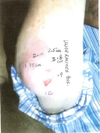
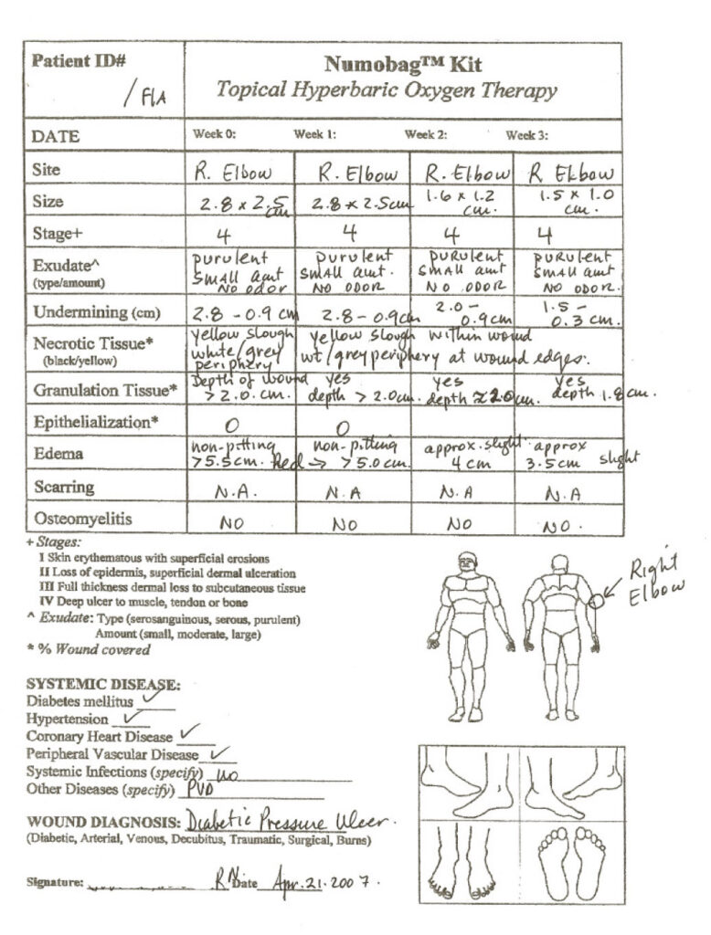
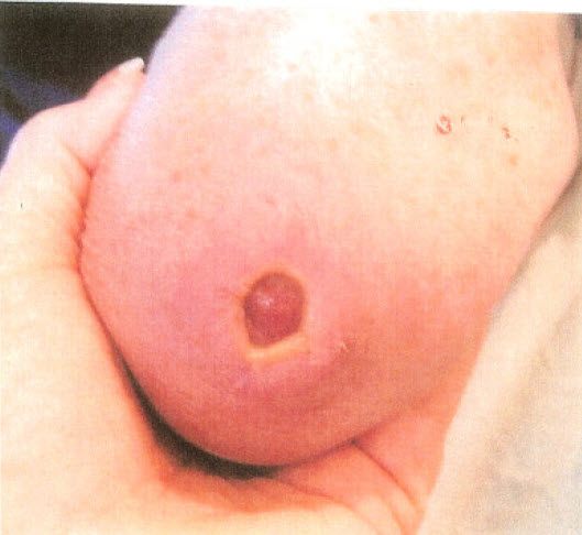
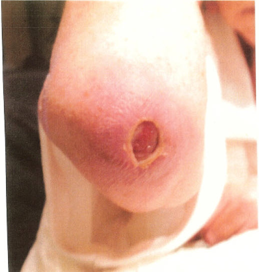
Wound opening: 2.8 x 2.5 cm
Depth: 2 cm
Exudate: Purulent, small amount, no odor
Edema and Erythema surrounding wound: 5.5 x 5.0 cm, warm, non-pitting edema. No granulation tissue present.
Necrotic tissue: Loosely adherent yellow slough within wound and white/grey tissue at periphery of wound opening. Edges are well defined, not attached to wound base, thickened and fibrotic.
Undermined tissue: 2.8 cm – 0.9 cm
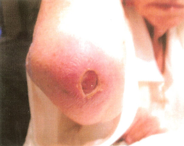
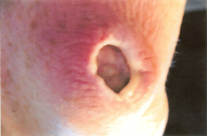
Wound opening: 2.8 x 2.5 cm
Depth: 2 cm
Exudate: Purulent, small amount, no odor
Edema and Erythema surrounding wound: Non-pitting edema extending >5.5 cm around wound with peripheral tissue induration, warm.
Necrotic tissue: Loosely adherent yellow slough within wound and white/grey tissue at periphery of wound opening. Edges are well defined, not attached to wound base, thickened and fibrotic.
Undermined tissue: 2.8 cm – 0.9 cm
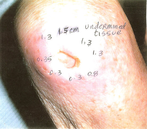
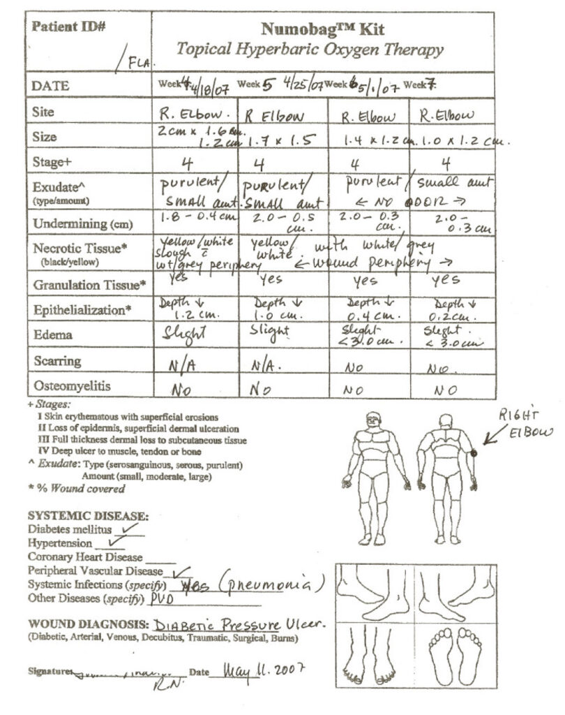
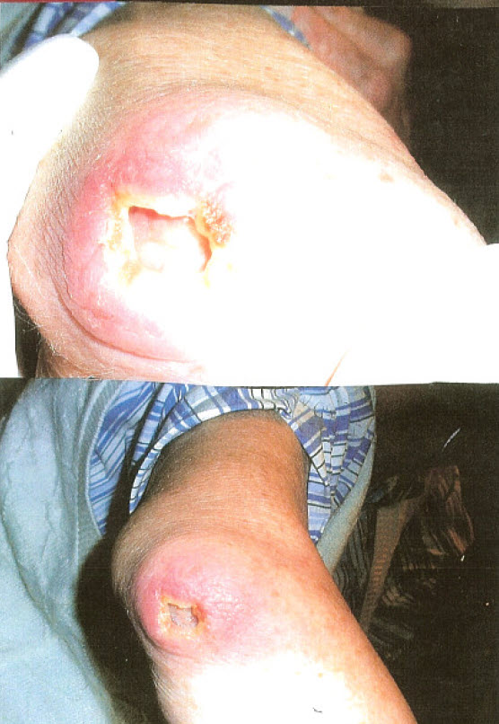
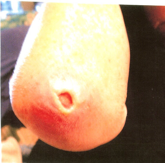
Wound opening: 1.5 x 1.0 cm
Depth: 1.8 cm
Exudate: Purulent, small amount, no odor
Edema and Erythema: 3.5 cm around wound, slight
Necrotic tissue: White/grey tissue at periphery of wound opening
Undermined tissue: 1.5 – 0.3 cm. Granulation tissue present at wound bed.
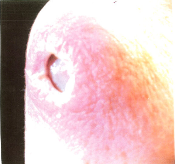
Wound opening: 2.0 x 1.2 cm
Depth: 1.2 cm
Exudate: Purulent, small amount, no odor
Edema and erythema: 4.0 cm around wound; slight
Necrotic tissue: White/gray tissue at periphery of wound opening
Undermined tissue: 1.8 – 4 cm. Granulation tissue present at wound bed.
Wound opening: 1.4 x 1.2 cm
Depth: 1 cm
Exudate: Purulent, small amount, no odor
Edema and Erythema surrounding wound: <3 cm around wound, slight
Necrotic tissue: White/gray at periphery of wound opening.
Undermined tissue: 2.0 – .5 cm. Granulation tissue present at wound bed.
Wound opening: 1.4 x 1.2 cm
Depth: 0.4 cm
Exudate: Purulent, small amount, no odor
Edema and Erythema: <3 cm, around wound, slight.
Necrotic tissue: White/gray at periphery of wound opening
Undermined tissue: 2.0 – 0.3 cm. Granulation tissue present at wound bed.
Wound opening: 1.4 x 1.2 cm
Depth: 0.4 cm
Exudate: Purulent, small amount, no odor
Edema and Erythema: <3 cm, around wound, slight.
Necrotic tissue: White/gray at periphery of wound opening
Undermined tissue: 2.0 – 0.3 cm. Granulation tissue present at wound bed.
Wound opening: 1.4 x 1.2 cm
Depth: 0.4 cm
Exudate: Purulent, small amount, no odor
Edema and Erythema: <3 cm, around wound, slight.
Necrotic tissue: White/gray at periphery of wound opening
Undermined tissue: 2.0 – 0.3 cm. Granulation tissue present at wound bed.
Wound opening: 1.0 x 1.2 cm
Depth: 0.2 cm
Exudate: Purulent, small amount, no odor
Edema and Erythema: <3 cm, around wound, slight.
Necrotic tissue: White/gray at periphery of wound opening
Undermined tissue: 2.0 – 0.3 cm. Granulation tissue present at wound bed.
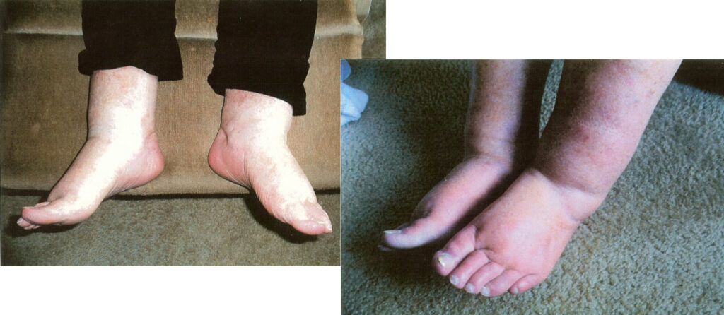
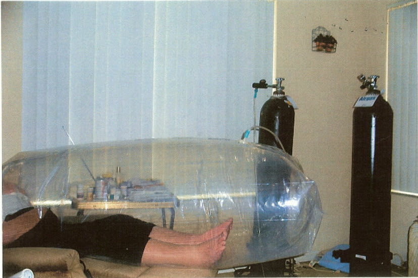
Right: Patient’s feet at the start of Numobag® treatments.